Microscopy with Lukey
One morning while Lukey and Kenzie were with us last week, Lukey and I decided to spend some time with my microscope. Kenzie popped in and out of the room a few times but spent most of the morning in Gee's office playing with her dollhouse as Gee worked. Kenzie and I had a few microscopy sessions together in the past but today was Lukey's turn. Kenzie was welcome to join us but she preferred to stick close to Gee and her dollhouse.
My desk is set up with all sorts of monitors for different tasks sort of resembling my own little Command Center and one of these monitors would be used to display what was under the microscope. On this morning, we had one of my Sony cameras mounted on the trinocular of the microscope. I used the binocular eyepieces for setting up each specimen. Lukey viewed what the camera was seeing on a 32 inch monitor over my desk. The view through the binocular eyepieces is far crisper than what is seen on the big monitor but this arrangement allowed us to view comfortably without the need for swapping places. Additionally, since I was the only one using the binocular eyepieces, we had no need keep adjusting focus and the pupillary distance of the binocular eyepieces whenever we swapped places. Being connected to this large overhead monitor made things much easier. For photos and video, I had a wireless remote connected to my camera.
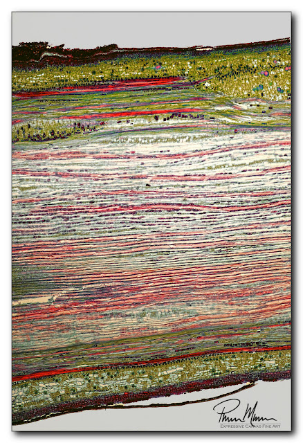
This first photo, below, shows a basswood specimen. My plan was to start by explaining that there is a distinct difference between cells of plants and cells of mammals. Lukey immediately exhibited some signs of losing interest likely because it was too much like school. I quickly explained that he's getting an introduction to science that he will need to know in middle school and high school so, when they start teaching this stuff, he'll be able to say, "Hey! I've seen this stuff before!" He liked the idea of getting a jump on this as opposed to most of his classmates. This thought seemed to bring him back to listening and engaging in what we were doing.
So, as I said, this first photo is a section of basswood. I pointed out the shape of the cells... being primarily rectangular and being lined up and organized. I also increased the magnification a bit more to point out the nucleus of each cell....
Here is a better photo showing an accurate size comparison between my hair and Lukey's hair...
I had a difficult time getting both hairs to lay flat in the photo that includes both hairs. This photo, above, was shot before I had used a drop of water and a coverslip to better control the two hairs so the perfectionist in me isn't happy with the quality of the image. Honestly, I wasn't going to include this photo but I decided to include it solely for the size comparison between Lukey's hair and my hair.
After talking about the forensic potential of examining hair under the microscope, I went outside to retrieve some standing water from our golf hole in the backyard. I wanted to compare our filtered tap water with this dirty standing water.
On the positive side, our double-filtered house tap water was completely devoid of anything to see under the microscope at magnifications up to 400x. That made me happy but there was nothing interesting to see under the microscope so we moved to the standing water from outside.
I was skeptical whether we would find anything in our standing water sample because it was below freezing outside all night long. I knew that there isn't much pond life in frozen water and especially frozen water that is devoid of plant life (food for the pond life). It was worth a shot anyway.
As you will see in the video, below, we did find some life in this small frigid standing water sample. There was some debris and some algae or plant-life. Then I found a small cell that was moving around between some debris and algae (or plant-life). At first I had trouble identifying it but I quickly realized it was a Testate Amoeba. Because this sample was so cold and rather devoid of other life making this a rather harsh environment, the amoeba had created its own little shell around itself for protection. As I was watching this amoeba, I noticed that the nucleus doubled... then the newer nucleus split from the amoeba as a smaller little amoeba... then it did that again... and again... and again. Rather than binary fission reproduction where the amoeba splits completely in half, we had captured multiple fission reproduction. I excitedly showed this to Lukey but I don't think he fully grasped what we were watching. It was a first for me though.
Then, another well formed cell passed through my view. This cell was green with a clear protective shell or bubble around it and it had a very well formed round shape. At first I thought it might be Volvox because it was so round and so well-formed but I quickly ruled that out. This was another Testate Amoeba called Arcella. I followed this amoeba hoping capture some other interesting action like we had seen in the previous Testate Amoeba but nothing happened. In the video, I also added a clip of pseudo-DIC microscopy (differential interference contrast). This makes it a little easier to see some structure of the cell.
Then the video shows some algae and then that video clip of the salt water drying to crystallized salt that I had mentioned above. That turned out nice and crisp. Focus was a bit difficult for the moving amoebae because they kept moving to different depths in the sample. So, I had to track in three dimensions. Still, the video is decent. I just wish I could have focused a bit more accurately.
NOTE: If you are wondering why you hear no sound, it is because the video has no sound. I deleted the sound because it was just background noise and some unrelated chatting in the room. Maybe I should have added some sort of background noise soundtrack like a babbling brook or similar.
Naturally, this relatively short session with the microscope sparked my interest in capturing far better images and videos of various specimens. Time is the biggest issue. My health is the priority and poor health cuts into available time. My to-do list is longer than ever too so hobbies tend to get pushed to the back-burner. And, I have far too many interests and hobbies!
Lukey and I had a good time looking at all these specimens under the microscope. I hope to share more of this with him in the future too!


Comments
Post a Comment